 RFI™, and the presumption of the existence of the Aura, is based upon the principle that all mental activity is electromagnetic. While some medical theorists argue that mental activity is chemical, because of the chemical neurotransmitters involved, the fact is that neurotransmitters are created only when electrical impulses induce a voltage in a neuron which exceeds its firing threshold. In addition, EEG (electroencephalogram) technology shows that mental activity can be effectively analyzed and monitored entirely by electromagnetic principles.
In electrical engineering, it is an established principle that all electrical currents produce surrounding electromagnetic (EM) fields. Accordingly, our psychological and emotional activities are sent throughout the body as electrical impulses, radiating electromagnetic fields outside the body, which are characteristic of the mental activity which generated them. Indeed, even the mere movement of atoms will produce a quantifiable electromagnetic field. This is most clearly observed in the case of charged radioisotopes, but even neutral atoms and molecules will emit a field through valence motion of the charged substructure particles. As proven by Russian Tesla technology experiments in the 1950s, this phenomenon causes prokaryotic organisms to produce an external electromagnetic field, which consists of the combined EM emissions from millions of molecules in the organism.
Most researchers in this field acknowledge that complex biological organisms such as humans also produce electrofield frequencies in much the same way, except that in addition to the molecular contribution, the nervous system plays a significant role. The nerves and nerve synapses carry electrical currents as large as 55 millivolts for neuron firings, and sometimes as large as 3 volts for motor impulses (to move the muscles), which radiate much larger electrical fields in accordance with electrofield physics. All of these radiating fields are the very tangible essence and definition of the human Aura.
RFI™ technology began as an experiment to develop a scientific basis for the clinical use of Aura imaging such as Kirlian photography, and to legitimize Aura imaging in general. While there are some highly effective and sophisticated Aura imaging devices on the market, there has previously been no empirical rationale for interpreting Aura colors to analyze psychological or physical health conditions.
To develop a system of interpretation suitable for clinical use, ITEM began with the scientific premise that color is frequency. ITEM was then able to calculate the resonant frequencies of actual color as light energy, and created mathematical formulations which translate all detectible electromagnetic frequencies into their corresponding colors. In this way, bioenergy colors can be detected by identifying the precise frequency of the bioenergy field in a specific location, and using the formulas to determine the actual color of the bioenergy.
The hypothesis of initial experiments conducted by ITEM was that if Aura photography and visualization was accurate, then Aura cameras and psychics would “visually” see the exact same colors as the objective Aura frequency colors scientifically identified by RFI™. In a controlled preliminary test of 10 reputable “psychic aura seers,” paired with 10 separate imaging clients, RFI™ verified the same colors as the psychics saw, all 10 times. In other experiments, 10 Russian “extrasenses” – who are certified specially trained bioenergy workers with medical education – tested RFI™ detection by willfully projecting specific colors in their Aura. In double-blind studies, the extrasenses would announce the color they intended to project, and RFI™ frequency data verified the extrasense’s color 100% of the time.
In January of 1995, ITEM gave a formal presentation of Resonant Field Imaging™ to the department heads of Moscow Bauman State Technical University, in Russia. The scientific rationale for proper interpretation of RFI™ images was discussed in detail, and the scientists of various disciplines offered comments and constructive criticism. Based upon the debate at this meeting, ITEM has developed technical principles which enhance the legitimacy and clinical usefulness of RFI™ Aura Imaging. ITEM continues this research today, and licensed users of RFI™ receive updates of any new advancements.
Scientists from the Russian Association of Human Ecology and Energyinformatic Science believe that the electromagnetic properties of the Aura detected by Kirlian and RFI™ methods may be only symptoms, or traces, of a deeper underlying cause. This cause is believed to be bioenergy in the form of torsion fields. At this time, all available data and research suggest that electromagnetic analysis through RFI™ is the most economical and tangible way to measure the secondary effects of torsion fields, without sacrificing accuracy or precision.
Many scientists and doctors have found it particularly intriguing that a technology can analyze psychological and emotional states. During ten years of research in this field, ITEM has found that emotion itself is essentially an energy reaction to a perception. First, one has a psychological perception of oneself and one’s environment. This perception, being a mental process, induces characteristic electrical impulses in the brain, which can affect related parasympathetic nerves or endocrine glands (e.g., a perception of fear stimulates the adrenal glands via the brain). Since the brain is neurologically connected to the entire human body, these electrical impulses may travel throughout the central, sympathetic and parasympathetic nervous system, creating the characteristic electrical fields.
Current research further suggests that certain levels of bioelectrical Aura fields are caused by more physical events in a biological organism. Biological activity such as autonomic responses initiate cellular and electrochemical changes, thereby creating an environment thermodynamically favorable to the conversion of metabolic kinetic energy into electromagnetic energy. In this process, localized bioenergy “complexes” to form a dynamic field that differentiates according to the neurological information that stimulated it. Since the muscles and skin are no barrier for such electromagnetic energy, the bioenergy field can and does radiate outside of the organism to become what we call the Aura.
According to the above biophysics, information about the “health” status of a complex organism is available from the coordination of sub-cellular energies, much the same as in a prokaryote. Any decrease in homeostasis, including parasitic infection, genetic mutation, arterial constriction, etc., will theoretically produce a corresponding modification in the bioelectrical field. RFI™ is an initial attempt to quantify and objectively interpret such modifications.
The accuracy of the RFI™ formulas and the color interpretations are tested extensively before each generation of prototypes is made available for our Scientific Investigators to test and use for their own analysis.
At one particular health conference where ITEM was testing and introducing RFI™, the resulting color Aura image may have played a role in helping to alert a woman of a life-threatening condition. A middle-aged woman requested a health-level RFI™ reading, and ITEM’s technician on duty discovered a “large red band” across the upper thorax region of the client. The technician, suspecting that an excessive concentration of red material energy could potentially be breast cancer, asked if she had ever had a mammogram, to which the woman responded that she had not. The technician advised that she may want to in the near future, emphasizing that this was not any type of diagnosis, but merely a logical precautionary suggestion. A few months later, the woman wrote ITEM and praised RFI™ technology for helping to identify a small malignant tumor that was successfully removed in time. She concluded the letter by stating: “RFI™ is a scientific instrument which may have helped to save my life, and I would recommend all alternative therapists use RFI™ as a supplement to their practice.” (This reference is provided only as an indication of the possible benefits of RFI™, and in no way implies that RFI™ can diagnose such conditions or guarantees you will have such results – see Disclaimer below).
The RFI™ package is the product of exhaustive research in the field of electrophysics and psychobiology. Whether you are a medical doctor, chiropractor, physical therapist, psychotherapist, alternative health practitioner, or just someone interested in exploring the new frontiers of science, we are confident that you will find RFI™ an interesting and useful tool in your endeavors.
There are 3 basic types of Aura devices on the market. The first category is “true Kirlian” photography devices. These use the original Aura technology as discovered by Russian scientist Seymon Davidovich Kirlian in the 1930s. Kirlian photography is the practice of placing the hand on an electrical plate, with a piece of photo paper between the hand and the plate. When the plate releases an electrical shock voltage, the Aura of the hand creates light exposure on the plate. The resulting image of the shapes of the hand’s Aura are amazing, and have been related to health conditions through principles of reflexology.
The disadvantages of true Kirlian photography are numerous: (1) it requires a real dark room like a professional photography laboratory, (2) it cannot reveal any information about psychological or mental energies, (3) it only shows the fingertips or at best the whole hand, (4) the electrical impulse generated by the Kirlian photography equipment, by definition, alter and distort the true shape of the Aura, by adding unnecessary voltage, at a pulse or frequency which cannot avoid influencing the natural energy information signals from the original Aura. The “Kirlian” class of devices generally cost between $1,000 and $3,000.
The second category of Aura devices are advertised as “Kirlian,” but are actually electrofield devices. Devices in the electrofield category do not use any direct photography, but use a sophisticated indirect system. First the client places their hands on the electroplates, which give an electrical pulse to amplify the natural aura for electrofield detection. Then, the computer detects and analyzes the resulting electrical biofield, and attempts to determine what color the Aura MIGHT look like, IF the device could actually see the Aura. Finally, the computer superimposes a cloud of that selected color onto the Polaroid photo for viewing. The resulting photos are stunning, and are certainly excellent proof of the existence of the Aura.
The disadvantages of this electrofield method are also numerous: (1) the resulting cloudy overlapping images can not be correlated to specific organs or neurological systems, and their distance from the body (essential to interpretation) cannot be determined from the photograph, (2) they make no distinction between the health level of the Aura and the emotional level of the Aura, and thus offer only vague metaphysical interpretations about a person’s spiritual state or mystical abilities, (3) these photographs usually only show the upper body, and exclude the hands, which often contain the most important information, (4) the electrical impulse necessarily distorts and contaminates the original Aura, so a technical analysis and interpretation cannot be made, (5) they do not show any numbers in scientific units, so the Aura images can not be verified or interpreted with the aid of any other available medical or scientific data. The “electrofield” class of Aura devices generally cost between $10,000 and $20,000.
The third category of Aura devices, which may be called “electro-meridian” devices, is an intelligent attempt to remedy the problems of interpretation inherent in the “electrofield” devices. First, instead of the electroplates giving an electrical impulse to amplify the Aura, they passively read the electrical activity at the fingertips, and use principles of meridians, acupuncture points and reflexology to determine where in the body the bioenergies detected in the fingers came from. A computer then calculates what colors those energies MIGHT look like IF it could see them, and superimposes the colors onto the Polaroid photograph. The result is an excellent photograph, with a more rational scientific basis, but it still has numerous disadvantages.
The disadvantages of “electro-meridian” imaging systems are: (1) the colors on the photograph describe biological conditions inside the physical body, but are artificially displayed as if outside the body, so the resulting photograph shows no information whatsoever as to the actual Aura field configuration either inside OR outside of the body, (2) they offer no distinction between the health and emotional levels of the Aura, (3) they do not show any numbers in scientific units, so the Aura images can not be verified or interpreted with the aid of any other available medical or scientific data, (4) they fail to detect or show any bioenergy emanating from the muscles, cells, parasympathetic or sympathetic nerves, or brain waves, all of which constitute the real human external biofield which defines the Aura. The “electro-meridian” devices generally cost between $2,000 and $5,000.
Resonant Field Imaging™ is not Kirlian photography. RFI™ is unique, and different from all of the above three categories of devices. It was engineered as a solution to all of the disadvantages of the other Aura systems.
Resonant Field Imaging™ can only be categorized as an “external electrofield imaging” technology. Since RFI™ is a passive measurement system, it does not cause any distortion or contamination of the Aura field, and gives a true, real-time reading of the original Aura field in its natural state. It literally measures the identity of the detected energy according to its characteristic frequency, and makes this frequency available to the user as data in standard international scientific units.
RFI™ has several unique advantages which none of the other systems can offer:
(1) It can be used to detect and analyze all existing energy fields, including the Auras of humans, objects, plants and animals, as well as ambient floating fields related to brain waves or even paranormal phenomena.
(2) The Aura image created by the software program shows clearly defined regions of the Aura, and distinguishes between the Health Level and Psychological (Emotional) Level of the Aura. It also clearly shows the identity and color of bioenergy in each of the 7 major Chakras. Chakras are bioenergy vortexes located along the spinal column, which contain energy-information related to corresponding endocrine glands.
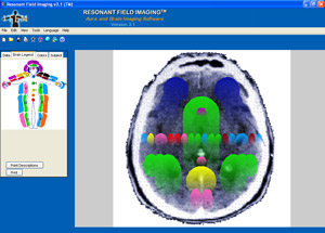
(3) It automatically generates medically and technically correct images of the human Brain, in full color, clearly revealing which color bioenergies are in specific regions and centers of the Brain.
(4) It prints out detailed interpretations of both the multi-level Aura and the Brain scan. The interpretations are based upon fundamental empirical science, and take into account the difference between Health Level bioenergies, Psychological (Emotional) Level bioenergies, and brain waves. Most importantly, the interpretations include explanations and detailed analysis of the significance of the identified bioenergies in the context of the regions where they are detected, and are therefore clinically meaningful and useful to practicing professionals, although they are generic by design because the system is not meant for diagnostic purposes.
The disadvantages and limitations of RFI™ are:
(1) It does not produce actual photographs, so it is necessary to understand (or explain) the scientific basis for its truth, accuracy and reliability, instead of placing reliance upon the appearance of photographs. To overcome this disadvantage, we developed the Technical Manual, to make you an expert in the scientific principles involved, and teach you how to express yourself effectively as a serious professional in this growing field.
(2) The software program cannot automatically generate images for objects, plants, animals, and ambient energy fields, because they are unpredictable in shape, and have wide variations in the number of levels of their Aura fields. To compensate for this limitation, we included the “Point / Object Analysis” function, which allows you to type one frequency number at a time into the computer, to obtain the color of the bioenergy you detected, as well as the Health Level or Psychological Level interpretation of that color. In addition, we also include a feature that allows you to import any picture that you choose, and specify the locations where you would like to take frequency measurements. This way, you can create completely unlimited full-color charts of anything you can imagine, by simply looking up the colors on the computer, and drawing the colors by hand. The Technical Manual gives you the expertise to explore unusual Auras and energy fields on your own, and develop your own customized interpretations.
Accordingly, Resonant Field Imaging™ is an ideal supplement to any Aura imaging devices which you may already own or wish to purchase, and the only viable choice for medical or scientific professionals who wish to use serious Aura imaging technology in clinical practice or in the laboratory. While other highly limited devices cost between $2,000 and $20,000, Resonant Field Imaging™ costs only $895.
All conventional brain imaging technologies, such as the CAT scan and PET scan, measure the properties of the brain indirectly, by first using a sensor to detect X-rays or radiation passing through the brain, compiling the numerical data, and then using a computer to construct a visual image of the brain, where the shapes and colors are superimposed onto the picture. There are two major disadvantages of using CAT and PET scans:
(1) It is necessary to subject the patient to X-rays passing through the head (CAT), or to inject the patient with a radioactive substance (PET), which limits how often a scan can be performed.
(2) The CAT scan only shows the shape, size and density of conditions in the brain. The PET scan only shows the intensity of activity in specific regions of the brain. While both are essential and beneficial in modern medicine, neither of them can reveal the causative factors of objects and events detected in the brain. Furthermore, both technologies are unable to provide information about what is happening in parts of the brain which have a normal appearance, or are inactive.
While the complex and integrated areas of the brain are too close together to read without expensive hospital type equipment, and it is too dangerous to place electrical probes into each area of the brain to obtain frequency readings, nature has solved this problem for us. Since electrical impulses from the brain are transmitted and radiate throughout the central, autonomic and parasympathetic nervous system, each area of our body to which the nerves extend radiate the brain signals which originated in the corresponding connected area of the brain. As a result, the human body becomes a macrocosm for the small scale of the complex brain, and serves as an accessible magnified window into the intricacies of the brain.
The RFI™ system generates a detailed, full color Brain scan image, by actually tracing the energies from regions of the human bioenergy field, back through the connecting neurological systems, and into the medically correct corresponding regions and organs of the human Brain. Just like the CAT and PET scans, the “RFI™ scan” accurately measures conditions and events in the brain indirectly, by first using the electrical field sensor to detect electromagnetic radiation originating in the brain, and then using a computer to compile the numerical data, and automatically construct a visual image of the brain.
While RFI™ can not be used to diagnose any particular diseases in the brain, it does have three major advantages:
 (1) RFI™ does not subject the patient to any radiation or unnatural substances which could have any side effects. Instead, it passively detects and analyzes the electrofield bioenergies which naturally emanate from the Brain. As a result, RFI™ Brain scans can be conducted as often as desired.
(2) RFI™ reveals, identifies and analyzes all bioenergies which are present in ALL major regions of the brain, regardless of whether they have a normal structure, and regardless of whether they are active or not. Therefore, RFI™ is able to give an enormous amount of information about structures of the brain which cannot be analyzed using the CAT or PET scans.
(3) By identifying the bioenergies which are present in specific regions and organs of the brain, RFI™ gives a more complete picture of the events, situations and conditions in the brain. It reveals which parts of the brain are effecting each other through the exchange of energy-information, and reveals the primary function or role of each part of the brain in a system of physical or mental illness. In addition, it is able to reveal exactly how the brain is influencing health conditions, and how health conditions and the patient’s psychology are physically affecting the brain.
Therefore, RFI™ brain scans are the ultimate tool in Health Psychology and Psychosomatic Medicine, and are a perfect supplement to conventional medical technologies.
![]()
![]()
Introduction to Resonant Field Imaging™ | Scientific Details | RFI™ Video | RFI™ Testimonials | Frequently Asked Questions | RFI™ Technical Manual | RFI™ Research | Locations of RFI™ Use | RFI™ Tutorial | Existing RFI™ Users Page | RFI™ Article | Your RFI™ System Includes | Order RFI™
RFI™ is an experimental process that is continuously under investigation by ITEM. ITEM uses its own research and the research of RFI™ Investigators to evaluate, improve and modify the process. The RFI™ system and its components are not intended for the diagnosis, cure, mitigation or treatment of any disease nor any other medical condition, and is not intended to affect the structure or any biological function of the body. RFI™ is strictly an “analysis tool” that may be used to identify psychological and physiological energy-information. This information can then be used in various ways depending on the training and experience of the investigator or practitioner.

Brain Optimization & Synchronization System™
- revolutionizing the way we think about energy and balance
© 2026 www.item-bioenergy.com. All rights reserved.